
Cutaneous Leiomyoma of the Leg: A Case Report and Literature Review
Authors
Files
Abstract
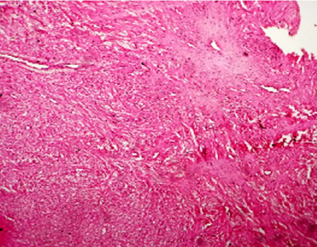
Cutaneous leiomyoma is a rare benign smooth muscle tumor that can be mistaken for other nodular cutaneous lesions. We present the case of a 27-year-old female graduate who developed a swelling on her left leg over a period of 2 years. Initially painless, the lesion became painful a year later, with pain exacerbated by heat. The lump measured 1 x 1 x 1 cm, was tender, and not warm. Initially diagnosed clinically as an epidermal inclusion cyst, she underwent excision biopsy revealing leiomyoma upon histological examination. Subsequent to excision, all symptoms resolved completely, with no recurrence noted. Hence, it is crucial to consider cutaneous leiomyoma as a potential differential diagnosis for such cutaneous lesions.
References
REFERENCES
Rowland F, Call C, Mujtaba B, Amini B, Wang WL. Calcified leiomyoma of the deltoid: pathophysiology and imaging review. Skeletal Radiol. 2019; 48(4):625-628. Doi: 10.1007/s00256-018-3053-y DOI: https://doi.org/10.1007/s00256-018-3053-y
Nessenius F, Zucal I, Allmann JK, Spreitzer S, Marti R. Incidental deep soft tissue leiomyoma of the groin – a case report and comprehensive review of literature. Journal of Surgical Case Reports. 2024; 1: 1–7 DOI: https://doi.org/10.1093/jscr/rjae020
Agulló Pérez AD, Resano Abárzuza MA, Córdoba Iturriagagoitia A, Aisa Rivera G, A. Patiño García A, Yanguas Bayona JI. Cutaneous leiomyomas: a clinicopathological and epidemiological review. An. Sist. Sanit. Navar. 2021; 44 (2): 163-176 DOI: https://doi.org/10.23938/ASSN.0914
Kilitci A, Elmas Ö F. Cutaneous Smooth Muscle Tumors: A Clinico pathological Study Focusing on the Under-Recognized Histological Features. Turk Patoloji Derg.2020; 36:126-134 DOI: https://doi.org/10.5146/tjpath.2019.01485
Ghanadan A, Abbasi A, Kamyab Hesari K. Cutaneous leiomyoma: Novel histologic findings for classification and diagnosis. Acta Med Iran.2013; 51:19-24.
Stout AP. Solitary Cutaneous and Subcutaneous Leiomyoma. The American Journal of Cancer. 1937; 29 (3):435–469. https://doi.org/10.1158/ajc.1937.435 DOI: https://doi.org/10.1158/ajc.1937.435
Kulkarni AR, Haase SC, Chung KC. Leiomyoma of the hand. HAND 2009; 4:145–149 DOI 10.1007/s11552-008-9143-x DOI: https://doi.org/10.1007/s11552-008-9143-x
Cinq E, Maruccia M, Malzone G, Malpassini F, Soda G, Drudi FM. A large vascular leiomyoma of the leg. Journal of Ultrasound.2012; 15:121-123.
Malhotra P, Walia H, Singh A, Ramesh V. Leiomyoma cutis: A clinicopathological series of 37 cases Indian J Dermatol.2010; 55:337-41. DOI: https://doi.org/10.4103/0019-5154.74535
Yokoyama R, Hashimoto H, Daimaru Y, Enjoji M. Superficial leiomyomas. A Clinicopathologic study of 34 cases. Acta Pathol Jpn. 1987; 37:1415-22 DOI: https://doi.org/10.1111/j.1440-1827.1987.tb02263.x
Alam NA, Barclay E, Rowan AJ, Tyrer JP, Calonje E, Marek S, Kelsell D et al. Clinical Features of Multiple Cutaneous and Uterine Leiomyomatosis: An Underdiagnosed Tumor Syndrome. Arch Dermatol. 2005; 141(2):199–206. doi:10.1001/archderm.141.2.199
Hachisuga T, Hashimoto H, Enjoji M. Angioleiomyoma: a Clinicopathologic reappraisal of 562 cases. Cancer 1984; 54:126e30. DOI: https://doi.org/10.1002/1097-0142(19840701)54:1<126::AID-CNCR2820540125>3.0.CO;2-F
Alam NA, Barclay E, Rowan AJ, Tyrer JP, Calonje E, Marek S, Kelsell D et al. Clinical Features of Multiple Cutaneous and Uterine Leiomyomatosis: An Underdiagnosed Tumor Syndrome. Arch Dermatol. 2005; 141(2):199–206. doi:10.1001/archderm.141.2.199 DOI: https://doi.org/10.1001/archderm.141.2.199
Cinq E, Maruccia M, Malzone G, Malpassini F, Soda G, Drudi FM. A large vascular leiomyoma of the leg. Journal of Ultrasound. 2012; 15:121-123. DOI: https://doi.org/10.1016/j.jus.2011.10.007